Explore CAMRI’s Advanced MRI Capabilities
At CAMRI, we combine cutting-edge MRI technology with expertise to deliver precise imaging for researchers, referrers and patients. Discover how CAMRI sets the standard for advanced diagnostics and research.
Why Choose CAMRI?
Precision Imaging
Precise imaging for accurate diagnosis and research.
Tailored Solutions
Dedicated solutions for researchers and medical specialists.
Clinical Expertise
Highly skilled and experienced MRI Technologists.
Reliable Results
Reliable results with expert reporting and support.
CAMRI's Expertise
Our services are continuously being updated. Please contact us if you have specific requests.
Cardiovascular
Our experienced team can offer high quality imaging of:
Paediatric and adult congenital heart disease
Adult acquired disease including both ischaemic and nonischaemic cardiomyopathies
Infiltrative myocardial disease assessment
Myocardial mapping and cardiac iron overload assessment with Ferriscan analysis.
Stress perfusion service
Cardiovascular assessment of aortic pathologies and peripheral arterial disease (PAD)
Imaging of patients with CIEDs using optimised sequences for metal artifact reduction (AL)
Neurology
The surgical planning for brain lesions can often be enhanced with specialist brain MRI sequences.We offer a comprehensive range of advanced imaging techniques, including:
Spectroscopy
- Single voxel
- 2D CSI (Chemical Shift Imaging)
- 3D CSIPerfusion and diffusion-weighted imaging
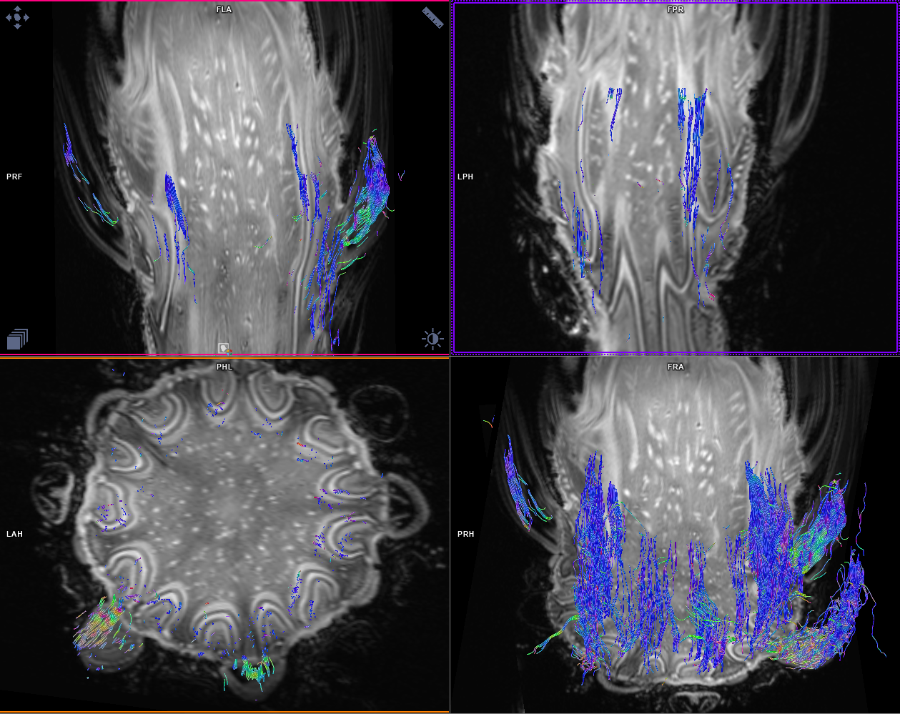
Diffusion tensor imaging (DTI) / fibre tracking
Arterial spin labelling (ASL)
Susceptibility-weighted imaging (SWI)
Functional MRI (fMRI)
- Motor tasks
- Language tasks
Advanced Liver
MR Elastography is a technology for non-invasive assessment of tissue stiffness.
MRE combines magnetic resonance imaging with low-frequency mechanical vibrations. The stiffness of tissues in the body will influence how the vibration waves can pass through them. This can be used to produce a map called an elastogram that measures the tissue stiffness.
MRE is most often used to detect stiffening of the liver caused by scarring (fibrosis) or inflammation and fat infiltration in chronic liver disease.
CAMRI has partnered with Resoundant to provide MRE measurements of tissue stiffness as part of the MRI examination, and to help evaluate some of the next generation of MRE technology.
Advantages of MRE Over Liver Biopsy
Unlike the traditional test for liver fibrosis which uses a needle to collect a sample of liver tissue (a biopsy), MRE as part of a full MRI examination offers several advantages:
MRE is more sensitive for detecting early fibrosis than other imaging methods.
MRE is a more comfortable, safer alternative to liver biopsy.
MRE images the entire liver, unlike a biopsy that evaluates just the small area of liver tissue.
MRE is more accessible for obese patients than some other imaging methods.
Liver MRE at CAMRI can be included as part of a full clinical MRI exam of the liver which also assesses liver morphology and liver fat content using ‘LiverLab’.MRE is not just limited to the liver, and researchers at CAMRI have also evaluated it to assess tissue mechanical properties in heart and brain.
FerriScan®
Measurement of liver iron concentration is critical in managing patients with potentially life-threatening iron overload conditions. CAMRI has partnered with Resonance Health to provide a quantitative and validated MRI assessment of tissue iron concentrations using FerriScan®.
FerriScan is globally recognised as the gold standard in the measurement of liver iron overload and is used internationally in over 30 countries. It provides a quality-assured measurement and has regulatory clearance in the United States, Europe, and Australia.
At CAMRI we offer liver iron quantification with liver fat assessment.
For patients at risk of iron-induced heart damage, FerriScan is available as a dual service with cardiac and liver iron quantification, including additional cardiac function assessment.
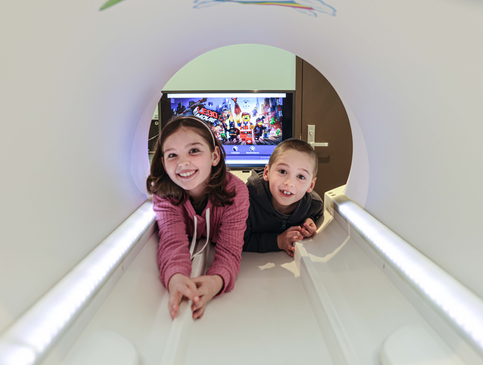
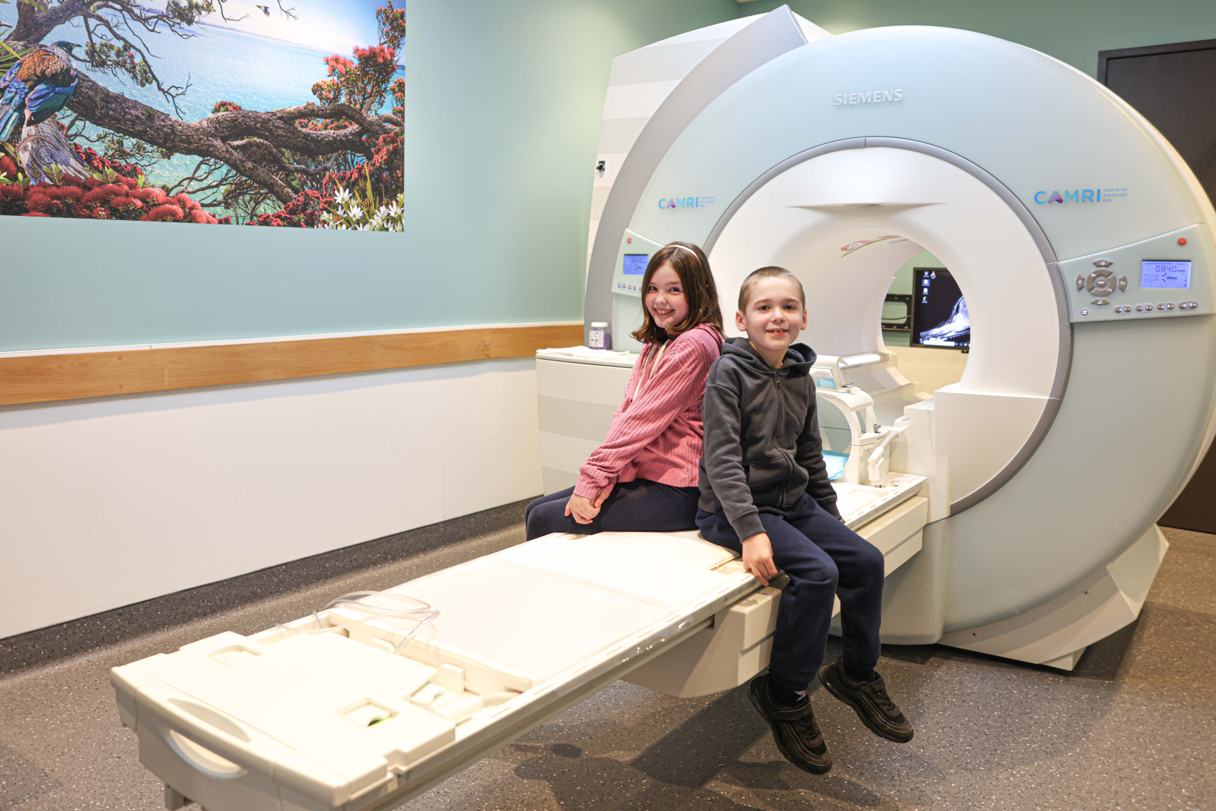
Paediatrics and Fetal
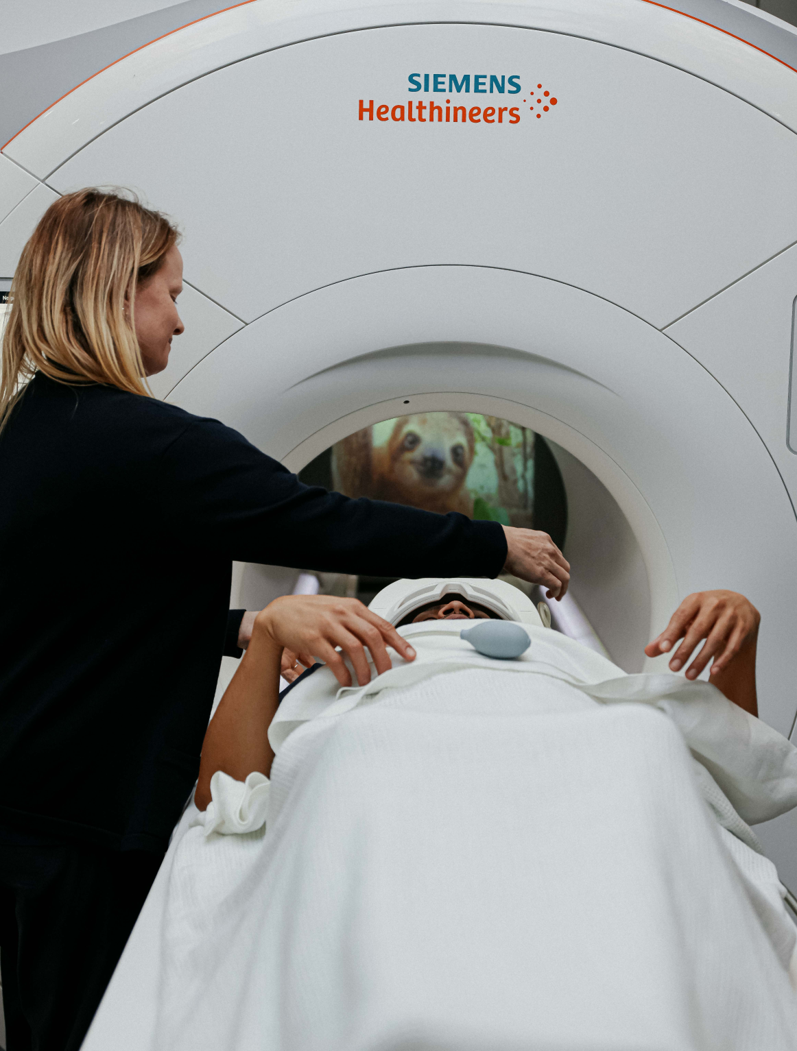
Asking a child to lie completely still inside a big, noisy scanner for 30–45 minutes can be frightening. Even the smallest amount of movement during an imaging sequence will degrade the images, making a competent diagnosis difficult.
If needed, children have the ability to practice their MRI in our mock scanner first to become comfortable with an MRI scanner.
Others may require general anaesthesia and CAMRI is able to offer these facilities to enable the best images to be acquired.
Specialties We Support
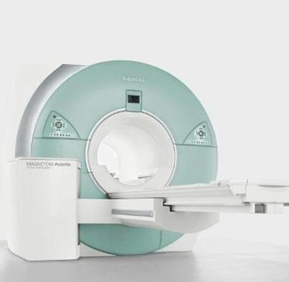
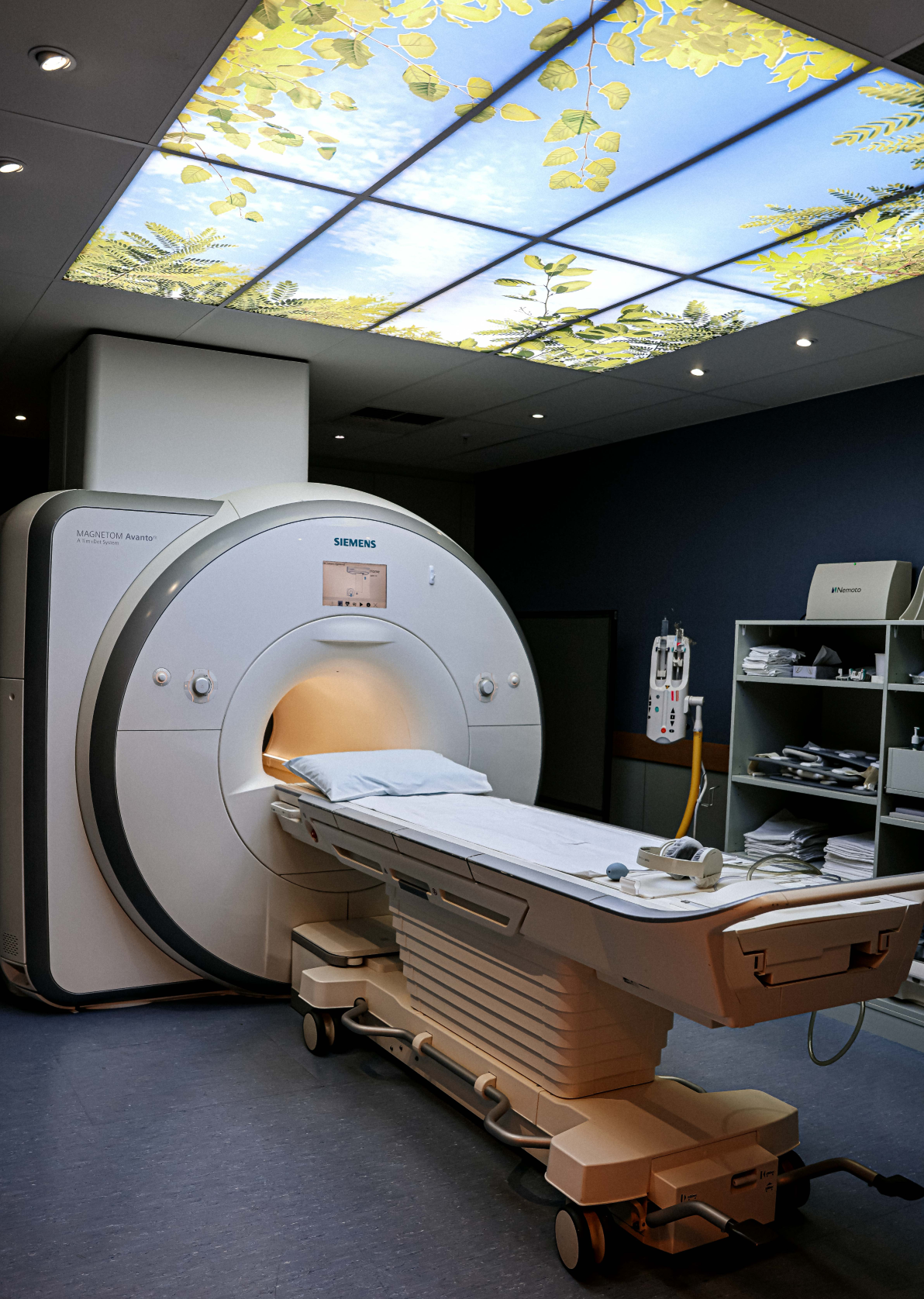
Our Technology
Our advanced MRI machines and tools
deliver unparalleled imaging quality.

"The mock MRI machine & experience is without a doubt the best thing for anxious kids & I'm sure adults too. Everyone was so friendly, helpful & understanding right from reception through to the radiologist. She felt so good she was able to get this done. Thank you for offering this invaluable service."
Begin Your Journey with CAMRI Today
Partner with us through referrals, schedule your imaging needs, or discuss your specific requirements with our expert team.


Paediatric
Asking a child to lie completely still inside a big, noisy scanner for 30–45 minutes can be frightening. Even the smallest amount of movement during an imaging sequence will degrade the images, making a competent diagnosis difficult.
If needed, children have the ability to practice their MRI in our mock scanner first to become comfortable with an MRI scanner.
Others may require general anaesthesia and CAMRI is able to offer these facilities to enable the best images to be acquired.
Child Entertainment
For children that are comfortable and relaxed in the MRI scanner, they can watch Netflix movies or TV shows, or they can listen to the music of their choice.
General Anaesthesia
The Centre for Advanced MRI (CAMRI) is one of the few private MRI facilities in New Zealand fully equipped with general anaesthesia and resuscitation equipment.
Our team of anaesthetists, technicians and recovery nurses are fully registered, and have expertise in paediatric, cardiac and other high risk patients. Their extensive experience enables us to offer the best possible care for you or your child.
MR Elastography (MRE)
MR Elastography is the non-invasive assessment of variations in tissue stiffness.
Liver fibrosis is an excessive accumulation of extracellular matrix proteins including collagen that occurs in most types of chronic liver diseases.
Advanced liver fibrosis results in cirrhosis, liver failure, and portal hypertension, where liver transplantation may be the only treatment option.
Diagnosis traditionally requires liver biopsy, and there is an acknowledged need for accurate, less invasive methods to detect and monitor chronic liver disease. These challenges have led to increasing interest in MR Elastography as a clinical and research tool.
The principle: by applying mechanical waves, MRI can now be used to non-invasively assess variations in tissue stiffness.
Hence, MR Elastography offers:
- A new MRI-based biomarker for characterising tissue non-invasively
- A new approach for improved treatment decisions, especially in liver fibrosis
Liver (Advanced)
MR Elastography is a technology for non-invasive assessment of tissue stiffness.
MRE combines magnetic resonance imaging with low-frequency mechanical vibrations. The stiffness of tissues in the body will influence how the vibration waves can pass through them. This can be used to produce a map called an elastogram that measures the tissue stiffness.
MRE is most often used to detect stiffening of the liver caused by scarring (fibrosis) or inflammation and fat infiltration in chronic liver disease.
CAMRI has partnered with Resoundant to provide MRE measurements of tissue stiffness as part of the MRI examination, and to help evaluate some of the next generation of MRE technology.
Advantages of MRE Over Liver Biopsy
Unlike the traditional test for liver fibrosis which uses a needle to collect a sample of liver tissue (a biopsy), MRE as part of a full MRI examination offers several advantages:
- MRE is more sensitive for detecting early fibrosis than other imaging methods.
- MRE is a more comfortable, safer alternative to liver biopsy.
- MRE images the entire liver, unlike a biopsy that evaluates just the small area of liver tissue.
- MRE is more accessible for obese patients than some other imaging methods.
Liver MRE at CAMRI can be included as part of a full clinical MRI exam of the liver which also assesses liver morphology and liver fat content using ‘LiverLab’.
MRE is not just limited to the liver, and researchers at CAMRI have also evaluated it to assess tissue mechanical properties in heart and brain.
Vascular
MR Elastography is a technology for non-invasive assessment of tissue stiffness.
MRE combines magnetic resonance imaging with low-frequency mechanical vibrations. The stiffness of tissues in the body will influence how the vibration waves can pass through them. This can be used to produce a map called an elastogram that measures the tissue stiffness.
MRE is most often used to detect stiffening of the liver caused by scarring (fibrosis) or inflammation and fat infiltration in chronic liver disease.
CAMRI has partnered with Resoundant to provide MRE measurements of tissue stiffness as part of the MRI examination, and to help evaluate some of the next generation of MRE technology.
Advantages of MRE
Unlike the traditional test for liver fibrosis which uses a needle to collect a sample of liver tissue (a biopsy), MRE as part of a full MRI examination offers several advantages:
- MRE is more sensitive for detecting early fibrosis than other imaging methods.
- MRE is a more comfortable, safer alternative to liver biopsy.
- MRE images the entire liver, unlike a biopsy that evaluates just the small area of liver tissue.
- MRE is more accessible for obese patients, than some other imaging methods.
Liver MRE at CAMRI can be included as part of a full clinical MRI exam of the liver which also assesses liver morphology and liver fat content using ‘LiverLab’.
MRE is not just limited to the liver, and researchers at CAMRI have also evaluated it to assess tissue mechanical properties in heart and brain.
Neurological
The surgical planning for brain lesions can often be enhanced with specialist brain MRI sequences.
We offer a comprehensive range of advanced imaging techniques, including:
- Spectroscopy
- Single voxel
- 2D CSI (Chemical Shift Imaging)
- 3D CSI
- Perfusion and diffusion-weighted imaging
- Diffusion tensor imaging (DTI) / fibre tracking
- Arterial spin labelling (ASL)
- Susceptibility-weighted imaging (SWI)
- Functional MRI (fMRI)
- Motor tasks
- Language tasks
FerriScan®
Measurement of liver iron concentration is critical in managing patients with potentially life-threatening iron overload conditions. CAMRI has partnered with Resonance Health to provide a quantitative and validated MRI assessment of tissue iron concentrations using FerriScan®.
FerriScan is globally recognised as the gold standard in the measurement of liver iron overload and is used internationally in over 30 countries. It provides a quality-assured measurement and has regulatory clearance in the United States, Europe, and Australia.
At CAMRI we offer liver iron quantification with liver fat assessment.
For patients at risk of iron-induced heart damage, FerriScan is available as a dual service with cardiac and liver iron quantification, including additional cardiac function assessment.
Cardiac
The Centre has a specific expertise in the following areas:
- Congenital heart disease
- Ventricular function in the context of cardiomyopathy and myocardial viability
- Pericardial disease
- Flow imaging
Ventricular function is assessed using advanced 3D software, which provides detailed measurements including:
- Cardiac mass
- Ventricular volumes
- Ejection fractions